Optical Coherence Tomography Applications
Ultra-high resolution Optical Coherence Tomography (UHR-OCT) is an invaluable tool enabling micron scale cross sectional or 3D imaging of the subject under investigation. Cross sectional or 3D imaging is relevant for various applications, ranging from analysis of tissue in medical applications to visualization of sub-micron structures in manufacturing.
High resolution optical microscopy is an invaluable tool for a wide range of applications. Optical Coherence Tomography (OCT) enables cross sectional or 3D imaging of the subject under investigation in contrast to alternative microscopy techniques where only the surface or shallow layers of the subject can be investigated. Cross sectional or 3D imaging is relevant for various applications, ranging from analysis of tissue in medical applications to visualization of sub-micron structures in manufacturing.
The OCT principle for imaging was first demonstrated 1991 by Huang et and a very thorough description of principles and applications has been provided by Drexler and Fujimoto in Optical Coherence Tomography: Technology and Applications Over the last 20 years OCT has developed into being an essential imaging tool in ophthalmology with particular emphasis on detailed analysis of the retina and the surrounding tissue. The applications in OCT are, however, not limited to ophthalmology and an increasing amount of research is made in OCT outside ophthalmology field.
The super continuum lasers offer extreme optical bandwidths, excellent spatial coherence and high optical power density that are key parameters for UHR-OCT. The number of OCT applications using super continuum lasers light sources is increasing rapidly.
OCT BASICS
OCT is based on interferometry, where light reflected or scattered off the subject under investigation interfere with light from a reference arm. The light from both arms originate from the same light source, and hence the two beams will interfere if the path length difference of the two arms in the interferometer is within the coherence length of the optical signal. This coherence gating allows the detection system to discriminate between reflections from closely spaced reflectors, thus enabling high resolution imaging.
OCT basic principle
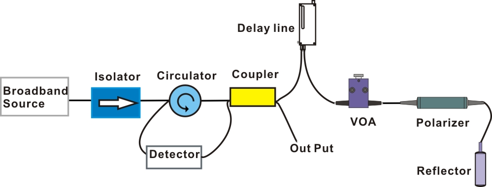
Figure 1: Schematic diagram of Optical Coherence Tomography
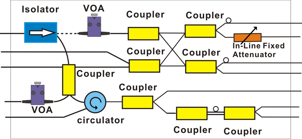
Figure 2: Schematic diagram module of Optical Coherence Tomography
The sensitivity of OCT can be very high, which enables even the weak signals originating from sub-surface reflections to be detected. In this way, cross sectional imaging of the subject under test can be realized in a way similar to ultrasound, but with a much better resolution, and image depths of several mm into tissue can be achieved.
DK Photonics offers a variety of passive components for building OCT systems, and we also manufacture integrated optical modules for OCT applications. DK Photonics could provide fused coupler, circulator, VOA, isolator, in-line reflector, collimator, WDM, in-line polarizer, and PBS with broadband working wavelength at 850 and 1064, 1310nm.
We also offer custom optical modules and subsystems by integrating multiple broadband components such as splitters, circulators, fixed and variable optical attenuators for various OCT applications These components are ideal in supporting the unique builds of the expanding global OCT interferometric systems. Contact DK Photonics regarding your requirements.
Click here to download the PDF file: 